
Ads by Google
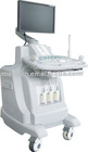

medical ultrasound Color Doppler system is intended for visualization of internal human organs and for medical diagnostic purposes only.
Clinic Applications
Abdominal, Vascular, OB/GYN, Cardiac, Urology, Breast, Small Parts, Musculoskeletal
Image Modes
B-Mode, M-Mode, Color Doppler (CFM), Doppler Spectrum Mode (PWD), Power Doppler Mode
Measurements & Calculations1) Basic B-mode: distance, area and circumference, A/B ratio, angle, ellipse, volume
2) Basic M-mode: velocity, heart rate, distance, slope, A/B ratio, time
3) Basic Doppler: peak velocity/frequency, time, mean velocity, heart rate, RI, PI, S/D ratio, flow volume, area
4) Basic Obstetrical Package: measurements include CRL, BPD, OFD, HC, TAD, AC, FL, APAD, FHR, user-defined measurement tables; calculations include: EFW, GA, EDD, HC/AC, FL/AC,FL/BPD, CI; reports cover: measurements averaging, tables and formulas, qualitative descriptions and pictogram display
5) Basic Gynecology Package: GYN and fertility protocol, size and volume of Ovaries, Uterus and Follicles, Cervix, Endometrial
6) Basic Cardiac Package: measurements include LVID, LVOT, LVPW, LA, LVET, LVPG for left ventricle and RVOT, RVD, RVET for right ventricle, cardiac output, cardiac index, FS, EF, heart rate; reports cover B-mode, M-mode and Doppler mode
7) Basic Urology Package: measurements include the volume of prostate, stepwise and residual Urine; reports cover: PSA/PSAD entry and calculation, qualitative descriptions and pictogram display
IMAGE FEATURES .
1) Display
Display Depth: up to 28 cm,
Probe dependent: Convex: 18 steps, Linear: 14 steps
Display Gray Levels: 256; continuous variable contrast and frame rate of up to 200 frames/sec
Display Format: B, 2B, 4B, M, B/D, B/M, B/C, B/C/MC, B/D, B/P, B/C/D
Image Orientation: Left/right B mode reversal and up/down image invert; 90, 1800 rotation
Magnification: Zoom with pan capability in real-time or freeze B, C and M mode
Annotation: Allows the user to annotate anywhere on the image with pre-defined annotation list and anatomical body marks
Screen Display: Display of all patient and exam related imaging parameters, and on-screen documentation of image parameters in single / dual display modes
2)3D & 4D Ultrasound
Offer freehand 3-D ultrasound with user training tools
Offer 3-D viewing and editing slices to remove unwanted tissue structure from arbitrary angles
Offer 3-D display in B-mode and Color Doppler simultaneously
4-D Picture forming capability
3) Image Review
CINE Review: Variable speed motion review and frame-by-frame review Storage only limited by internal memory of the system board
Standard: 1,024 B-Mode frame and 170 seconds M-Mode data
Standard: 520 color frames and 380 seconds Doppler Spectrum
Post Procession and measurements
4) Image Management
Storage only limited by hard disk of the system board
Standard: 160 GB hard disk drive for local image storing
CD-RW/DVD Drive as removable read, write, archive and storage, USB, S-Video, VGA
Standard: 2,000 Images w/ TIF format on CD
DICOM & PACs Compatible
Standard Configuration
1) Main unit, 15”LCD monitor, 3 probe connectors, 2 USB ports
2)3.5MHz Convex Array Probe, 7.5MHz Linear Probe,
Transducer Options:
Linear array: 5.0 MHz, 7.5 MHz
Curved array: 3.5 MHz
Trans-vaginal: 7.5 MHz
Phased array: 2.5 MHz
4D Transducer: Curved 3.5 MHz
Video printer
Free-hand 3D
DICOM,CW,Free Steering M,
Color Doppler